How could computer modelling be used for cancer treatment?
In the treatment of cancer, oncological imaging methods play a significant role, but it takes time and effort to interpret the images. Machine learning and artificial neural networks coud be used in the analysis of PET scans in the future.Published: 6.2.2025
Text: Oona Rainio
Picture: iStock
Editing: Viestintätoimisto Jokiranta Oy
Cancer is among the most frequent causes of death in the world. Globally, approximately 20 million new cancer cases are diagnosed every year, and the volume is expected to increase along with the ageing of the population and the rising standard of living. Various imaging methods play a significant role in the treatment of cancer as they can be used to locate and measure cancer tumours in the human body. They also facilitate the monitoring of a patient’s disease, its progress over time and response to different treatments.
One of these medical imaging methods is positron emission tomography (PET), which is based on the use of short-lived radioactive substances. For PET scanning, the patient is intravenously injected with a radioactive tracer, which will spread in the body through blood circulation. A commonly used tracer for oncological PET scanning is fluorodeoxyglucose (FDG), which contains fluorine-18, a radioactive isotope of fluorine with a half-life of about two hours. FDG also contains glucose, a source of energy for the human body.
Fluorodeoxyglucose accumulates in cancer tumours. This occurs because cancer cells divide in an uncontrolled manner and, therefore, have a higher demand for glucose than the surrounding healthy tissue. Due to its unstable composition, the radioactive FDG emits small particles known as positrons. The positron annihilation reactions result in gamma radiation, which can be recorded by means of a special PET camera. The scanning yields real-time information about the spreading of the tracer in the body. From PET images, doctors can see in which areas of the body the tracer is being accumulated and thereby locate cancer tumours.
Machine learning is helpful for image analysis
The analysis of PET scanning images is a laborious process. Typically, doctors denote cancer tumours by means of segmentation, that is, by drawing the outlines of the tumours in the scans, so as to facilitate the rapid comparison of changes occurring between two PET scannings. Various automated tools could help doctors in doing this job. It is much faster to simply check whether a cancer tumour is correctly segmented than to manually draw its location in the scan.
The automated segmentation of cancer tumours calls for machine learning. Machine learning refers to computer models that not only follow specific rules defined in advance but are also capable of independent learning. For the purposes of learning, the model is provided with a vast amount of training data and instructions on the desired outcome. The aim is for the model to first learn what it should do with the training data in order to achieve the desired outcome. Then, the model can be generalised to process other datasets apart from those used in training.
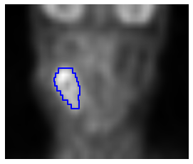

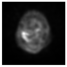
Fig. 1. A vertical cross-section of a three-dimensional fluorodeoxyglucose PET image of a head and neck cancer patient with the light area indicating high tracer accumulation. Cancer tumour segmentation denoted by the doctor is marked in blue.
Fig. 2. A horisontal cross-sectional image from the same patient, given to the convolutional neural network (CNN) for prediction.
Fig. 3. A segmentation mask drawn by the doctor for the horisontal cross-sectional image in Fig. 2 and given to the CNN for prediction. White pixels are cancerous and black pixels either healthy tissue or background.
Source: Joonas Liedes, Turku PET Centre, TYKS (Turku University Central Hospital).
A convolutional neural network is an efficient tool
A convolutional neural network (CNN) is a potential machine learning model that is specifically intended for the processing of images. Artificial neural networks are models comprised of millions of parameters and programmed to imitate the transfer of information in the neural cells of the human brain. In this context, convolution refers to a specific matrix calculation, through which the model is able to process the scans and save the information produced by adjacent pixels in the images.
A convolutional neural network can be programmed to predict a positive or negative value for each pixel in a given scan. In terms of oncological imaging, a positive pixel falls within the area of a cancer tumour whereas a negative pixel is not cancerous. The CNN model is then trained by providing it with hundreds or thousands of scans, together with scan-specific segmentation masks that contain the information about the correct pixel categorisation.
Accordingly, a convolutional neural network can be taught to recognise cancer tumours from PET scans on the basis of tracer accumulation, provided that there is enough data available with known correct categorisations for the model training. The model programming and data gathering processes are laborious, but the CNN will also learn to predict new scans. Once a sufficiently accurate CNN is developed for the recognition of a particular type of cancer, doctors can use it as a tool in their clininal work.
Oona Rainio earned her PhD in Mathematics from the University of Turku in autumn 2021. During the past three years, she has worked as a post-doctoral researcher at Turku PET Centre. Her research concerns the application of machine learning and statistical methods in the processing of medical scanning images.
Read more:
- https://www.duodecimlehti.fi/duo15553
- https://link.springer.com/article/10.1007/s13721-024-00483-0
- https://link.springer.com/article/10.1007/s42600-023-00314-7
- https://www.nature.com/articles/s41598-024-56706-x
- https://sites.utu.fi/intoimaging/en/